|
Cayman Chemical
recombinant human pad4  Recombinant Human Pad4, supplied by Cayman Chemical, used in various techniques. Bioz Stars score: 90/100, based on 1 PubMed citations. ZERO BIAS - scores, article reviews, protocol conditions and more https://www.bioz.com/product/recombinant+human+pad4+enzyme/pmc08560728-56-22-27?v=Cayman+Chemical Average 90 stars, based on 1 article reviews
recombinant human pad4 - by Bioz Stars,
2026-08
90/100 stars
|
Buy from Supplier |
|
Proteintech
pad4 enzyme  Pad4 Enzyme, supplied by Proteintech, used in various techniques. Bioz Stars score: 96/100, based on 1 PubMed citations. ZERO BIAS - scores, article reviews, protocol conditions and more https://www.bioz.com/product/recombinant+human+pad4+enzyme/bio_rxiv__2025__02__28__639952-55-5-33?v=Proteintech Average 96 stars, based on 1 article reviews
pad4 enzyme - by Bioz Stars,
2026-08
96/100 stars
|
Buy from Supplier |
|
ModiQuest
recombinant human pad2  Recombinant Human Pad2, supplied by ModiQuest, used in various techniques. Bioz Stars score: 90/100, based on 1 PubMed citations. ZERO BIAS - scores, article reviews, protocol conditions and more https://www.bioz.com/product/recombinant+human+pad4+enzyme/pmc04036085-68-0-8?v=ModiQuest Average 90 stars, based on 1 article reviews
recombinant human pad2 - by Bioz Stars,
2026-08
90/100 stars
|
Buy from Supplier |
|
Corning Life Sciences
high-binding elisa plates High Binding Elisa Plates, supplied by Corning Life Sciences, used in various techniques. Bioz Stars score: 90/100, based on 1 PubMed citations. ZERO BIAS - scores, article reviews, protocol conditions and more https://www.bioz.com/product/recombinant+human+pad4+enzyme/pm37778379-93-74-77?v=Corning+Life+Sciences Average 90 stars, based on 1 article reviews
high-binding elisa plates - by Bioz Stars,
2026-08
90/100 stars
|
Buy from Supplier |
|
Cayman Chemical
human recombinant histone h3 cayman #10263  Human Recombinant Histone H3 Cayman #10263, supplied by Cayman Chemical, used in various techniques. Bioz Stars score: 90/100, based on 1 PubMed citations. ZERO BIAS - scores, article reviews, protocol conditions and more https://www.bioz.com/product/recombinant+human+pad4+enzyme/pmc08722705-65-7-11?v=Cayman+Chemical Average 90 stars, based on 1 article reviews
human recombinant histone h3 cayman #10263 - by Bioz Stars,
2026-08
90/100 stars
|
Buy from Supplier |
|
Cayman Chemical
biotin-phenylglyoxal (pg  Biotin Phenylglyoxal (Pg, supplied by Cayman Chemical, used in various techniques. Bioz Stars score: 90/100, based on 1 PubMed citations. ZERO BIAS - scores, article reviews, protocol conditions and more https://www.bioz.com/product/recombinant+human+pad4+enzyme/pmc10326043-89-6-10?v=Cayman+Chemical Average 90 stars, based on 1 article reviews
biotin-phenylglyoxal (pg - by Bioz Stars,
2026-08
90/100 stars
|
Buy from Supplier |
|
Biomol GmbH
recombinant pad3 cay10786  Recombinant Pad3 Cay10786, supplied by Biomol GmbH, used in various techniques. Bioz Stars score: 90/100, based on 1 PubMed citations. ZERO BIAS - scores, article reviews, protocol conditions and more https://www.bioz.com/product/recombinant+human+pad4+enzyme/bio_rxiv__2025__03__27__645732-74-27-30?v=Biomol+GmbH Average 90 stars, based on 1 article reviews
recombinant pad3 cay10786 - by Bioz Stars,
2026-08
90/100 stars
|
Buy from Supplier |
|
Corning Life Sciences
rehydrated matrigel chamber  Rehydrated Matrigel Chamber, supplied by Corning Life Sciences, used in various techniques. Bioz Stars score: 90/100, based on 1 PubMed citations. ZERO BIAS - scores, article reviews, protocol conditions and more https://www.bioz.com/product/recombinant+human+pad4+enzyme/pmc08492346-194-51-53?v=Corning+Life+Sciences Average 90 stars, based on 1 article reviews
rehydrated matrigel chamber - by Bioz Stars,
2026-08
90/100 stars
|
Buy from Supplier |
|
MyBiosource Biotechnology
anti-ra33 assay, any isotype, Anti Ra33 Assay, Any Isotype,, supplied by MyBiosource Biotechnology, used in various techniques. Bioz Stars score: 90/100, based on 1 PubMed citations. ZERO BIAS - scores, article reviews, protocol conditions and more https://www.bioz.com/product/recombinant+human+pad4+enzyme/pm39815378-69-31-37?v=MyBiosource+Biotechnology Average 90 stars, based on 1 article reviews
anti-ra33 assay, any isotype, - by Bioz Stars,
2026-08
90/100 stars
|
Buy from Supplier |
|
Tecan Systems
elisa reader  Elisa Reader, supplied by Tecan Systems, used in various techniques. Bioz Stars score: 99/100, based on 1 PubMed citations. ZERO BIAS - scores, article reviews, protocol conditions and more https://www.bioz.com/product/recombinant+human+pad4+enzyme/pmc05440486-29-107-109?v=Tecan+Systems Average 99 stars, based on 1 article reviews
elisa reader - by Bioz Stars,
2026-08
99/100 stars
|
Buy from Supplier |
|
Proteintech
pvdf membrane Pvdf Membrane, supplied by Proteintech, used in various techniques. Bioz Stars score: 94/100, based on 1 PubMed citations. ZERO BIAS - scores, article reviews, protocol conditions and more https://www.bioz.com/product/recombinant+human+pad4+enzyme/bio_rxiv__2025__02__28__639952-55-24-33?v=Proteintech Average 94 stars, based on 1 article reviews
pvdf membrane - by Bioz Stars,
2026-08
94/100 stars
|
Buy from Supplier |
|
Avantor
hepes Hepes, supplied by Avantor, used in various techniques. Bioz Stars score: 90/100, based on 1 PubMed citations. ZERO BIAS - scores, article reviews, protocol conditions and more https://www.bioz.com/product/recombinant+human+pad4+enzyme/pm37683647-213-80-82?v=Avantor Average 90 stars, based on 1 article reviews
hepes - by Bioz Stars,
2026-08
90/100 stars
|
Buy from Supplier |
Image Search Results
Journal: Frontiers in Immunology
Article Title: Applicability of Small-Molecule Inhibitors in the Study of Peptidyl Arginine Deiminase 2 (PAD2) and PAD4
doi: 10.3389/fimmu.2021.716250
Figure Lengend Snippet: Inhibitory capacity of AFM-30a, GSK199, and BB-Cl-amidine on citrullination of fibrinogen. ELISA plates were coated with human fibrinogen. (A) Recombinant human PAD2 or (B) PAD4 were used for citrullination of fibrinogen in the presence of BB-Cl-amidine, AFM-30a, or GSK199 at various concentrations. DMSO was used as a vehicle control. Following a 2-h incubation, a monoclonal antibody (clone 20B2) recognizing a citrullinated epitope of fibrinogen was added to assess the extent of citrullination. PAD activity was normalized to that observed in the absence of inhibitors (=100%) and is expressed as the mean ± SD of duplicates.
Article Snippet: Hereafter, wells were incubated with 30 ng/ml of recombinant human PAD2 (cat. 10785, Cayman Chemicals, Ann Arbor, MI, USA) or 150 ng/ml
Techniques: Enzyme-linked Immunosorbent Assay, Recombinant, Incubation, Activity Assay
Journal: bioRxiv
Article Title: Citrullination of TDP-43 is a key post-translation modification associated with structural and functional changes and progressive pathology in TDP-43 mouse models and human proteinopathies
doi: 10.1101/2025.02.28.639952
Figure Lengend Snippet: a, The enzymatic reaction mediated by PADs converts arginine to citrulline. b, c, Western blot images of PAD2 and PAD4-mediated citrullination of H3 (b) and TDP-43 (c). In (c) arrows indicate the observed shift in TDP-43 molecular weight. d, Coomassie staining of PAD2 (bands 1, 4), PAD4 (bands 2, 6), unmodified TDP-43 (band 3), PAD2-mediated citR TDP-43 (band 5), and PAD4-mediated citR TDP-43 (band 7). e, Bar diagrams showing the MW shift (kDa) of TDP-43 protein following citrullination. f, Schematic representation of arginine epitopes positioned in the human TDP-43 protein sequence. Position of the 11 citrullinated arginine epitopes are indicated in red. g, Six out of eleven arginine epitopes susceptible to citrullination laid within common RX(X)R (red box) or RXG/RGGG (green box) motifs in TDP-43 sequence. h, MS/MS spectrum showing b- and y-ion coverage of modified citR83 peptide and extracted ion chromatograms (XICs) showed abundance and retention time of unmodified R83 vs. citR83 with PAD2 and PAD4 treatment (intact peptide monoisotopic m/z (+2) 719.8457, (+3) 480.2329). i, Retention time peaks for TDP-43 peptides surrounding the unmodified or modified R83 epitope and base peak m/z undergoing methionine (Met85) oxidation, citrullination or both showed reliable time separation.
Article Snippet: Upon citrullination with PAD2 or
Techniques: Western Blot, Molecular Weight, Staining, Sequencing, Tandem Mass Spectroscopy, Modification

Journal: bioRxiv
Article Title: Citrullination of TDP-43 is a key post-translation modification associated with structural and functional changes and progressive pathology in TDP-43 mouse models and human proteinopathies
doi: 10.1101/2025.02.28.639952
Figure Lengend Snippet: Spectra showing b- and y-ion coverage of citrullinated peptides, as well as XICs retention times for unmodified and citrullinated peptides treated with PAD2 and PAD4, corresponding to a - c, citR165 (intact peptide monoisotopic m/z (+2) 665.8214), d - f, citR191(intact peptide monoisotopic m/z (+2) 672.8469), g - i, citR268/272 (intact peptide monoisotopic m/z (+2) 728.3560), j - l, citR293 (intact peptide monoisotopic m/z (+2) 638.29).
Article Snippet: Upon citrullination with PAD2 or
Techniques:

Journal: bioRxiv
Article Title: Citrullination of TDP-43 is a key post-translation modification associated with structural and functional changes and progressive pathology in TDP-43 mouse models and human proteinopathies
doi: 10.1101/2025.02.28.639952
Figure Lengend Snippet: a, Schematic representation of seven polyclonal antibodies raised against the citrullinated arginine epitopes. Epitope positions within each TDP-43 functional domain is indicated. b, Antibody specificity was determined by western blot via PAD4-mediated citrullinated TDP-43 protein probed with citR83 (NLS), citR165 and citR191 (RRM1, RRM2), citR268/272, citR275 (CTD) antibodies. c, Immunohistochemical images of TAR4/4 and non-Tg littermates cortical tissue labeled against citR TDP-43 antibody panel. Increased neuronal expression of citR83, citR165, citR191, citR268/272 and citR275 in TAR4/4 tissue compared to Non-Tg tissue. Arrows indicate the citR TDP-43 morphologies observed with the citR268/272 and citR275 antibodies raised against TDP-43 C-terminal domain (arrows). Insets represent magnified neuronal citR TDP-43 morphological properties recognized from each antibody. d, Images of cortical tissue incubated with secondary antibody only - “no-primary” control. n = 3, scale bar is 50µm and 200 µm.
Article Snippet: Upon citrullination with PAD2 or
Techniques: Functional Assay, Western Blot, Immunohistochemical staining, Labeling, Expressing, Incubation, Control
Journal: bioRxiv
Article Title: Citrullination of TDP-43 is a key post-translation modification associated with structural and functional changes and progressive pathology in TDP-43 mouse models and human proteinopathies
doi: 10.1101/2025.02.28.639952
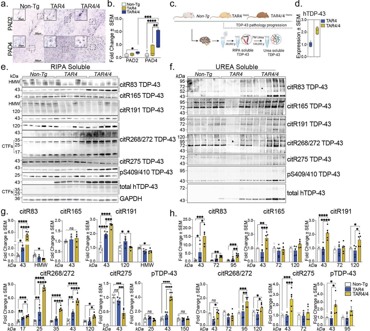
Figure Lengend Snippet: a, Immunohistochemical images of Non-Tg, TAR4 and TAR4/4 cortex labeled with PAD2 and PAD4 antibodies. b, Fold change of PAD2 and PAD4 (% area mean of the values ± SEM) normalized to the Non-Tg control. One-way or Two-way ANOVA, followed by Tukey’s post hoc multiple comparisons tests, n = 5, *p < 0.05, **p < 0.01, ***p < 0.001, ****p < 0.0001. c, Schematic overview of the TAR TDP-43 mouse model and the fractionation of cortical tissue into RIPA-soluble and (7 M) Urea-soluble fractions. d, The expression levels of the human TDP-43 in the TAR model. e, Cortical RIPA soluble and f, Urea soluble fraction analyzed by Western blotting and probed for citR TDP-43 antibody panel (citR83, 165, 191, 268/272, 275), pTDP-43 409/410 and total human TDP-43 protein. g, Quantification of citR TDP-43 43 kDa protein levels, proteolytic fragments (17, 25 & 35 kDa) and intermediate high molecular species (72, 98 & 120k Da) normalized to GAPDH in RIPA and h, Urea fraction normalized to the total protein loaded signal. Data represent the mean of the values ± SEM; One-way ANOVA, followed by Tukey’s or Šidák post hoc multiple comparisons tests, n = 5, *p < 0.05, **p < 0.01, ***p < 0.001, ****p < 0.0001.
Article Snippet: Upon citrullination with PAD2 or
Techniques: Immunohistochemical staining, Labeling, Control, Fractionation, Expressing, Western Blot
Journal: Journal of immunology (Baltimore, Md. : 1950)
Article Title: Citrullination alters immunomodulatory function of LL-37 essential for prevention of endotoxin-induced sepsis
doi: 10.4049/jimmunol.1303062
Figure Lengend Snippet: Human MDMs (A) and mouse RAW 264.7 (B) macrophages were stimulated with 10 ng/mL LPS in the presence of native or citrullinated LL-37 at the indicated concentrations (0.1–10 μg/mL). Citrullinated LL-37 was obtained by treatment of the native peptide with human PAD2 or PAD4 at 23.3 U/mg peptide). The level of TNF-α (A) and NO (B) in the culture supernatants was determined using ELISA or the Griess assay at 6 or 20 h post-stimulation, respectively. Since neither the LL-37 nor the PAD enzymes alone induced the release of NO or TNF-α, for the sake of clarity these controls are not shown in the figure. Data represent the mean ± SD of three independent experiments. ns, not significant; *, p<0.05; **, p<0.01; ***, p<0.001.
Article Snippet:
Techniques: Enzyme-linked Immunosorbent Assay, Griess Assay
Journal: Journal of thrombosis and haemostasis : JTH
Article Title: Quantification of citrullinated histones: Development of an improved assay to reliably quantify nucleosomal H3Cit in human plasma
doi: 10.1111/jth.15003
Figure Lengend Snippet: Recombinant nucleosomes are superior calibration standards for plasma-based enzyme-linked immunosorbent assays (ELISAs) compared to either recombinant or enzymatically modified histone proteins. A, In vitro citrullinated histone H3 calibration standard curves were prepared using two different lots of peptidyl-arginine deiminase 4 (PAD4) enzyme (2.5 U PAD4 per μg histone), with serial dilutions of each preparation tested by ELISA in parallel. The lots produce statistically distinct calibration curves; F(DFn, DFd) 133.3 (4,6), P < .0001. B, Recombinant H3R2,8,17Cit designer nucleosomes (dNucs), but not recombinant H3R2,8,17Cit histones, are recovered based on expected levels after direct dilution into 100% human plasma. C, ELISA standard curves for lots 1 and 2 of H3R2,8,17Cit dNucs display high inter-lot consistency. There is no statistical difference between the curves; F(DFn, DFd) 2.186 (4,34), P = .0915. D, ELISA standard curves for lots 2 and 3 of H3R2,8,17Cit dNucs (lot 1 was exhausted at the time of these experiments) also show no statistical difference between the curves; F(DFn, DFd) 2.004 (4,10), P = .1698
Article Snippet: Briefly, human recombinant PAD4 (Cayman #10500) and
Techniques: Recombinant, Clinical Proteomics, Modification, In Vitro, Enzyme-linked Immunosorbent Assay
![Citrullinated histone H3 (H3Cit)-DNA enzyme-linked immunosorbent assay (ELISA) performance metrics. A, Intra-assay variability. The same plasma sample in six replicates on the same plate showed an intra-assay coefficient of variation (CV) of 3.3%. B, Inter-assay variability. Four plasma samples (S1-S4) analyzed in duplicate on four different days showed an inter-assay CV of 7.4, 12.5, 6.2, and 6.5%, respectively, with a mean (standard deviation) inter-assay CV of 8.9% (2.9%). C, Dilution linearity. Two samples were spiked with H3R2,8,17Cit designer nucleosomes (dNucs) to 2000 ng/mL and serially diluted in assay buffer. Mean (standard deviation [SD]) recovery for dilutions within the working range of lower and upper limits of quantification was 88% (18%). D, Parallelism. Two samples with high endogenous H3Cit-DNA complex levels were serially diluted in assay buffer. Mean (SD) CV was 16.9% (3%). E, Recovery. Human plasma was prepared in different dilutions. Known concentrations of H3R2,8,17Cit dNucs were spiked into plasma diluted 1:1–1:8 to theoretical concentrations of 400 ng/mL. Recovery were all within 80%–120%, with a mean (SD) % recovery of 93.3% (10.4%). F, Selectivity. The assay detected H3R2,8,17Cit dNucs, but not unmodified recombinant nucleosomes. G, Analysis of plasma samples from 40 cancer patients and 30 healthy individuals ensured that the levels of H3Cit-DNA complexes in human plasma were within working range of the assay, and revealed significantly higher levels in cancer patients than in healthy individuals; median (interquartile range) 295.0 (151.5–489.1) versus 27.9 (9.9–78.9), P < .001](https://pub-med-central-images-cdn.bioz.com/pub_med_central_ids_ending_with_2705/pmc08722705/pmc08722705__nihms-1645894-f0004.jpg)
Journal: Journal of thrombosis and haemostasis : JTH
Article Title: Quantification of citrullinated histones: Development of an improved assay to reliably quantify nucleosomal H3Cit in human plasma
doi: 10.1111/jth.15003
Figure Lengend Snippet: Citrullinated histone H3 (H3Cit)-DNA enzyme-linked immunosorbent assay (ELISA) performance metrics. A, Intra-assay variability. The same plasma sample in six replicates on the same plate showed an intra-assay coefficient of variation (CV) of 3.3%. B, Inter-assay variability. Four plasma samples (S1-S4) analyzed in duplicate on four different days showed an inter-assay CV of 7.4, 12.5, 6.2, and 6.5%, respectively, with a mean (standard deviation) inter-assay CV of 8.9% (2.9%). C, Dilution linearity. Two samples were spiked with H3R2,8,17Cit designer nucleosomes (dNucs) to 2000 ng/mL and serially diluted in assay buffer. Mean (standard deviation [SD]) recovery for dilutions within the working range of lower and upper limits of quantification was 88% (18%). D, Parallelism. Two samples with high endogenous H3Cit-DNA complex levels were serially diluted in assay buffer. Mean (SD) CV was 16.9% (3%). E, Recovery. Human plasma was prepared in different dilutions. Known concentrations of H3R2,8,17Cit dNucs were spiked into plasma diluted 1:1–1:8 to theoretical concentrations of 400 ng/mL. Recovery were all within 80%–120%, with a mean (SD) % recovery of 93.3% (10.4%). F, Selectivity. The assay detected H3R2,8,17Cit dNucs, but not unmodified recombinant nucleosomes. G, Analysis of plasma samples from 40 cancer patients and 30 healthy individuals ensured that the levels of H3Cit-DNA complexes in human plasma were within working range of the assay, and revealed significantly higher levels in cancer patients than in healthy individuals; median (interquartile range) 295.0 (151.5–489.1) versus 27.9 (9.9–78.9), P < .001
Article Snippet: Briefly, human recombinant PAD4 (Cayman #10500) and
Techniques: Enzyme-linked Immunosorbent Assay, Intra Assay, Clinical Proteomics, Inter Assay, Standard Deviation, Recombinant
Journal: Frontiers in Immunology
Article Title: Citrullination of C1-inhibitor as a mechanism of impaired complement regulation in rheumatoid arthritis
doi: 10.3389/fimmu.2023.1203506
Figure Lengend Snippet: Confirmation of C1-INH citrullination by PAD4 and PAD2 using the PG-biotin probe on ELISA and Western blotting. C1-INH was incubated with PAD2 or PAD4 under reducing conditions and in the presence of 8 mM of CaCl 2 . Citrullination of C1-INH was confirmed using the biotin-PG probe at low pH and visualized in ELISA (A) and Western blotting (B) .
Article Snippet: Recombinant human PAD2 and PAD4 and
Techniques: Enzyme-linked Immunosorbent Assay, Western Blot, Incubation

Journal: Frontiers in Immunology
Article Title: Citrullination of C1-inhibitor as a mechanism of impaired complement regulation in rheumatoid arthritis
doi: 10.3389/fimmu.2023.1203506
Figure Lengend Snippet: ELISA confirming ACPA reactivity to citrullinated C1-INH. (A, B) RA patient samples were added to microtiter plates coated with native and PAD2-citrullinated (A) as well as PAD4-citrullinated (B) C1-INH. Autoantibody binding was detected using HRP-conjugated rabbit anti-human IgG and compared between ACPA-negative and ACPA-positive samples for PAD2-citrullinated (C, E) and PAD4-citrullinated C1-INH (D, F) for all RA patient samples (C, D) as well as for merely RF-negative samples (E, F) . Data are presented as median plus interquartile range, and significance was calculated using Mann–Whitney U and Kruskal–Wallis rank-sum tests. Citrullination of C1-INH using 100 U of the PAD enzymes per 1 mg of protein was confirmed using the biotin-PG probe visualized by Western blotting **p < 0.01, ***p < 0.001, ****p < 0.0001 (G) . ACPA, anti-citrullinated protein antibody; RA, rheumatoid arthritis; HRP, horseradish peroxidase; RF, rheumatoid factor.
Article Snippet: Recombinant human PAD2 and PAD4 and
Techniques: Enzyme-linked Immunosorbent Assay, Binding Assay, MANN-WHITNEY, Western Blot
Journal: bioRxiv
Article Title: Development of Motif-Specific Monoclonal Antibodies for Global Protein Citrullination Detection with Minimal Cross-Reactivity to Homocitrullination
doi: 10.1101/2025.03.27.645732
Figure Lengend Snippet: (A) Reactivity of motif-specific monoclonal antibodies 12A2 and 21F10 against in vitro citrullinated or homocitrullinated HeLa proteomes. (-), untreated HeLa lysate. (B) Reactivity of three commercially available anti-pan citrullination antibodies (07-377, ab100932, and MABN328 (F95)) against citrullinated and homocitrullinated HeLa lysates. (-), untreated HeLa lysate. (C) Reactivity of motif-specific monoclonal antibodies 12A2 and 21F10 toward HeLa proteomes treated with different PAD isozymes (PAD1, PAD2, PAD3, PAD4). (-), untreated HeLa lysate.
Article Snippet: For comparison of the citrullination patterns of different PAD isozymes, 200 μg of HeLa lysate (5 μg/μL) was diluted with 160 μL PAD buffer and incubated with
Techniques: Bioprocessing, In Vitro
Journal: JCI Insight
Article Title: DDR1-induced neutrophil extracellular traps drive pancreatic cancer metastasis
doi: 10.1172/jci.insight.146133
Figure Lengend Snippet: (A – D ) Human neutrophils were cocultured with DDR1 knockdown or reexpression of MDA-PATC 148 or BxPC-3 by Matrigel transwell chamber, with or without NADPH oxidase inhibitor, PDA4 inhibitor, NE inhibitor, and Dase I treatment for 18 hours. ( A ) NET structures were analyzed by immunofluorescence staining using DAPI (blue), anti-NE (red), and anti-histone H3 (green) mAbs. Scale bar : 50 μm. ( B ) The NET quantification is displayed as NET histone area (μm 2 ) per x40 field, 6 fields per group. ( C and D ) The number of invaded cells analyzed by immunofluorescence staining using DAPI and calculated based on the number of cells found in 6 fields per chamber. All the data are mean ± SD. n = 5, 3 independent experiments; 1-way ANOVA with Sidak post hoc testing. * P < 0.05; ** P < 0.01; *** P < 0.001.
Article Snippet: We suspended 2 × 10 5 cancer cells in 500 μL of serum-free DMEM medium (D0 medium) with or without 7rh, IgG, anti-CXCL5 antibody, BSA, human recombinant CXCL5, DNase I, NADPH oxidase inhibitor, apocynin, PAD4 inhibitor, Cl-amidine, and the neutrophil elastase inhibitor sivelestat and then added the cells to a rehydrated
Techniques: Knockdown, Immunofluorescence, Staining

Journal: JCI Insight
Article Title: DDR1-induced neutrophil extracellular traps drive pancreatic cancer metastasis
doi: 10.1172/jci.insight.146133
Figure Lengend Snippet: ( A – F ) Human neutrophils were cocultured with DDR1 knockdown or reexpression of MDA-PATC 148 or BxPC-3 by Matrigel transwell chamber, with or without anti-CXCL5 neutralized antibody or recombinant CXCL5 treatment, for 18 hours. ( A ) NET structures were analyzed by immunofluorescence staining using DAPI (blue), anti-NE (red), and anti–histone H3 (green) mAbs. Scale bar: 50 μm. ( B and C ) The NET quantification is displayed as NET histone area (μm 2 ) per field, 6 fields per group. ( D ) Cit-histone H3 expression were analyzed by western blotting. ( E and F ) The number of invaded cells were analyzed by immunofluorescence staining using DAPI and calculated based on the number of cells found in 6 fields per chamber. All the data are mean ± SD. n = 5, 3 independent experiments. ( B and E ) P values were analyzed by 1-way ANOVA with Sidak post hoc testing. *** P < 0.001. ( C and F ) P values were analyzed by unpaired 2-tailed Student’s t test. * P < 0.05; *** P < 0.001.
Article Snippet: We suspended 2 × 10 5 cancer cells in 500 μL of serum-free DMEM medium (D0 medium) with or without 7rh, IgG, anti-CXCL5 antibody, BSA, human recombinant CXCL5, DNase I, NADPH oxidase inhibitor, apocynin, PAD4 inhibitor, Cl-amidine, and the neutrophil elastase inhibitor sivelestat and then added the cells to a rehydrated
Techniques: Knockdown, Recombinant, Immunofluorescence, Staining, Expressing, Western Blot

Journal: JCI Insight
Article Title: DDR1-induced neutrophil extracellular traps drive pancreatic cancer metastasis
doi: 10.1172/jci.insight.146133
Figure Lengend Snippet: ( A and B ) MDA-PATC 148 cells were pretreated with 7rh for 30 minutes and then with collagen I for 3 hours. ( A ) Phospho-NF-κB P65, phospho-PKCθ, and phospho-SYK were analyzed by western blotting. ( B ) CXCL5 levels were analyzed by ELISA. Data are mean ± SD. n = 4, 3 independent experiments; 1-way ANOVA with Sidak post hoc testing. * P < 0.05; *** P < 0.001. ( C – E ) Human neutrophils were cocultured with MDA-PATC 148 and BxPC-3 cells by Matrigel transwell chamber for 18 hours. ( C ) NET structures were analyzed by immunofluorescence staining using DAPI (blue), anti-NE (red), and anti–histone H3 (green) mAbs. Scale bar: 50 μm. ( D ) The NET quantification is displayed as NET histone area (μm 2 ) per field, 6 fields per group. Data are mean ± SD. n = 6, 3 independent experiments; 1-way ANOVA with Sidak post hoc testing. *** P < 0.001. ( E ) Cit-histone H3 expression were analyzed by western blotting. ( F and G ) Mice were orthotopically injected with MDA-PATC 148 cells, with or without 3 mg/kg 7rh treatment for 9 weeks. ( F ) Liver metastasis was detected by immunofluorescence staining using DAPI (blue) and anti-CK19 (red) mAbs in liver section. Scale bar: 50 μm. The metastasis quantification is displayed as CK-19 positive signals/per x20 field, 6 fields per group. ( G ) Neutrophils infiltration was detected by immunofluorescence staining using DAPI (blue), anti-CK19 (red), and anti-Ly6G (green) mAbs in pancreas section. Scale bar: 50 μm. The Neutrophils infiltration quantification is displayed as Ly6G positive signals per x20 field, 6 fields per group. Data are mean ± SD. n = 5, unpaired 2-tailed Student’s t test. *** P < 0.001.
Article Snippet: We suspended 2 × 10 5 cancer cells in 500 μL of serum-free DMEM medium (D0 medium) with or without 7rh, IgG, anti-CXCL5 antibody, BSA, human recombinant CXCL5, DNase I, NADPH oxidase inhibitor, apocynin, PAD4 inhibitor, Cl-amidine, and the neutrophil elastase inhibitor sivelestat and then added the cells to a rehydrated
Techniques: Western Blot, Enzyme-linked Immunosorbent Assay, Immunofluorescence, Staining, Expressing, Injection
Journal: Immunologic Research
Article Title: Validation of an enzyme-linked immunosorbent assay for the quantification of citrullinated histone H3 as a marker for neutrophil extracellular traps in human plasma
doi: 10.1007/s12026-017-8905-3
Figure Lengend Snippet: Schematic of the H3Cit ELISA procedure. A Anti-histone biotin (the capture antibody) is coated to streptavidin pre-coated wells during the first incubation. Samples are pipetted into the wells and histones bind to the capture antibody during the second incubation. B After washing, anti-H3Cit is added to the wells, binding to immobilized H3Cit but not to histones H3 that are not citrullinated, during the third incubation. C In the fourth incubation, an HRP conjugated anti-rabbit antibody is added and binds to the anti-H3Cit, after which TMB is added for detection
Article Snippet: Phosphate buffered saline (PBS; Life Technologies, Cat. No. 14190-250), tween 20 (Sigma-Aldrich, Cat. No. A9418), rabbit polyclonal anti-histone H3 (citrulline R2 + R8 + R17) antibody (Abcam, Cat. No. AB5103), bovine serum albumin, BSA (Sigma-Aldrich, Cat. No. A9418), goat anti-rabbit IgG horseradish-peroxidase (HRP) conjugate (BioRad, Cat. No. 170-6515), 3,3′, 5,5′-tetramethylbenzidine (TMB) liquid substrate (Sigma-Aldrich, Cat. No. T0440), stop solution (Thermo Scientific, Cat. No. N600), Trizma base (Sigma-Aldrich, Cat. No. T1503), CaCl 2 (Sigma-Aldrich C1016), phenylmethylsulfonyl fluoride (PMSF) protease inhibitor (Life Technologies, Cat. No. 36978), dithiothreitol, DTT (Invitrogen, Cat. No. P2325), human recombinant PAD4 (Cayman Chemical, Cat. No. 10500), human recombinant histone H3 (Cayman Chemical, Cat. No. 10263),
Techniques: Enzyme-linked Immunosorbent Assay, Incubation, Binding Assay